Research

FLASH highlights the path to dynamic resilience in European healthcare financing

The EU-funded FLASH(opens in new window) project is exploring solutions to tackle the rigidity of healthcare financing models and their inability to adequately handle sudden, large-scale disruptions. A stark reminder is the COVID-19 pandemic. While European nations responded by injecting emergency funds and reallocating resources, these measures highlighted the need for flexibility. To help improve the preparedness, efficiency and equity of EU health systems, FLASH analyses three critical dimensions of flexibility.

Sudden shocks, care and health needs

One dimension focuses on the flexibility to absorb sudden shocks. The pandemic revealed that even well-performing health systems struggled with surges in demand, often resulting in postponed care for non-COVID-19 conditions. FLASH developed a resilience measure to assess how quickly hospital systems return to pre-crisis activity levels. Their analysis identifies three pillars of resilience: a robust workforce with effective interdisciplinary coordination; digital integration, including electronic health records and real-time monitoring; and the ability to learn from experience. The researchers conclude that hospital resilience cannot be achieved through isolated interventions but requires an integrated approach strengthening human, technological and organisational capacities simultaneously.

Another examines flexibility regarding where care is received. Ideally, patients should access high-quality care regardless of EU residence, yet barriers to cross-border mobility persist. FLASH investigates legal and economic obstacles, demonstrating that economic barriers can often be overcome with existing legislative frameworks. By proposing methods to harmonise cross-country payment systems and tariffs, the project suggests that facilitating patient mobility can actually increase social welfare, particularly in terms of border mobility and facilities’ capacity management.

A third addresses flexibility with respect to health needs, specifically tackling inequities in funding allocation. While co-payments reduce financial burdens on public payers, they often lead to unmet needs among disadvantaged populations. FLASH extends this analysis to the inter-regional level, investigating whether increased health needs in specific regions are matched by increased financial resources. The findings reveal significant heterogeneity across the EU. While Belgium and Spain see funding increase in line with needs, this association is weak in Czechia and Italy. By cataloguing resource allocation rules in nine countries, the project provides policymakers with a reference to reform existing rules governing the allocation of funding from the central to the local level.

The case of Catalonia

However, the push for flexibility and digitalisation carries its own risks, as highlighted in a recent FLASH (Flexible Approaches to Support Health through financing) study(opens in new window) in Catalonia. It evaluated the adoption and long-term use of the Catalan National Health System’s digital platform ‘MyHealth’, covering over 9.4 million individuals and 727 million health interactions between 2015 and 2023. The results showed that despite rapid expansion accelerated by the pandemic, access to digital health services remains deeply unequal. Engagement was significantly higher among women, Spanish nationals and higher-income individuals, while older adults, migrants and rural residents remained consistently underrepresented.

The conclusion reached is that digitalisation alone does not guarantee greater equity in healthcare access. As they strive for greater flexibility, EU health systems need to simultaneously address structural barriers such as digital literacy, linguistic and cultural accessibility, and infrastructure in disadvantaged areas. Ultimately, a truly flexible healthcare system is one that adapts not only to financial and logistical shocks, but also to the diverse needs of its entire population.

By building a network of procurement of innovation stakeholders, the EU-funded Procure4Health project is helping European healthcare systems move from reactive buying to a more demand-driven approach

Across Europe, healthcare faces the challenge of doing more with limited resources. “Procurement usually focuses on buying finished products,” says project coordinator Carlos Larrañeta Gómez-Caminero, from the Andalucian Public Foundation for Progress and Health(opens in new window) (website in Spanish) in Spain. “We wanted to approach it the other way round. Defining our challenges and then going out to the market to find solutions.”

Flipping the model

Procure4Health(opens in new window) set out to change how European public health systems adopt innovation. Instead of adapting to solutions already on the market, the project encouraged public health buyers to identify their needs first and then invite developers to respond.

Procure4Health brought together 33 partners from 15 countries, covering health and social care procurers and supporting organisations at local, regional and European level, all testing new ways of bringing innovation into healthcare.

One of the project’s main tasks was to help buyers pin down their procurement needs. Partners worked in groups on topics such as digital health, sustainability, precision medicine and integrated care, alongside work on value and impact. Together, they defined the challenges common to all countries, decided which ones to prioritise and tested them with suppliers through open market consultations, following the EAFIP(opens in new window) methodology.

The project also funded 29 cascade training opportunities helping spread experience across Europe and supported twinning partnerships designed to share knowledge and build capacity, alongside a detailed action plan(opens in new window) produced for health organisations new to the approach. Additionally, insights and takeaways from the project were published and brought together in a book(opens in new window).

A concrete result

One of Procure4Health’s key outcomes is THERESA PCP, a follow-on EU project which brings together hospitals and research organisations from Belgium, Estonia, the Netherlands, Poland and Spain to develop on-site systems for treating hospital wastewater and removing some of healthcare’s most hazardous pollutants.

Currently, there is no single process that handles antibiotics, cytostatic drugs and antibiotic-resistant bacteria altogether. “An open market consultation showed that solutions existed for individual pollutants, but not for all of them together,” notes Larrañeta. A pre-commercial procurement approach allowed health systems to develop and test a more complete solution. “Seeing this shared challenge developed during the project turn into a funded PCP was a real achievement,” he adds.

Cross-border cooperation

The project also changed how health buyers collaborate across Europe. In Belgium and France, central purchasing bodies involved in the project formed a new partnership, allowing them to work together more effectively on innovative solutions. For smaller or more fragmented health systems, this kind of joint approach can make procurement of innovation easier and more economically viable.

Procure4Health also highlighted the importance of value-based procurement, especially for healthcare providers trying to judge whether a new solution is worth adopting in practice. This remained one of the project’s key follow-on areas, as partners continue to work on procurement criteria that go beyond price alone.

The road ahead

Although the project ended in May 2025, work hasn’t stopped for Procure4Health. Partners continue to focus on procurement criteria related to sustainability and value, while developing follow-on proposals. The project also published a policy paper(opens in new window) that makes recommendations and proposes practical measures to strengthen understanding, build capacity and scale up innovation procurement in healthcare across Europe.

But for Larrañeta, this is about more than procurement. “Almost 25 % of clinicians’ weekly activity is devoted to administrative tasks,” he notes. Reducing that burden could free up more time for clinicians to focus on patients.

Cambridge scientists have grown miniature circuits in the lab that mimic how the brain and spinal cord connect up, which underlies our movements. They used this model to show how damage to these connections previously considered ‘irreversible’ could, in fact, be reversible

As we develop and grow from embryo to fetus to infant, our nerve cells (neurons) form connections, allowing information to be transmitted between the brain and the spinal cord. A key component of each neuron is the axon – the nerve fibre ‘cable’ that transmits information to other neurons to activate muscle contractions. 

At some point, we lose the ability to grow axons in the central nervous system, or this ability is at least greatly impaired or slowed down. This means that damage to the brain and spinal cord becomes permanent, leading to devastating disabilities, such as the inability to grasp or walk. This is often the case for traumatic spinal cord injury and can be a feature of many neurological diseases, including motor neurone disease or multiple sclerosis. 

In 2021, Dr András Lakatos and colleagues at the University of Cambridge developed ‘mini brains’ using human patient-derived stem cells – special cells that have the potential to develop into most human cell types – which they guided to grow into pea-sized brain ‘organoids’. These organoids were 3D models that resemble parts of the human cerebral cortex. The team used these to demonstrate molecular problems in motor neurone disease and potential ways to prevent them.

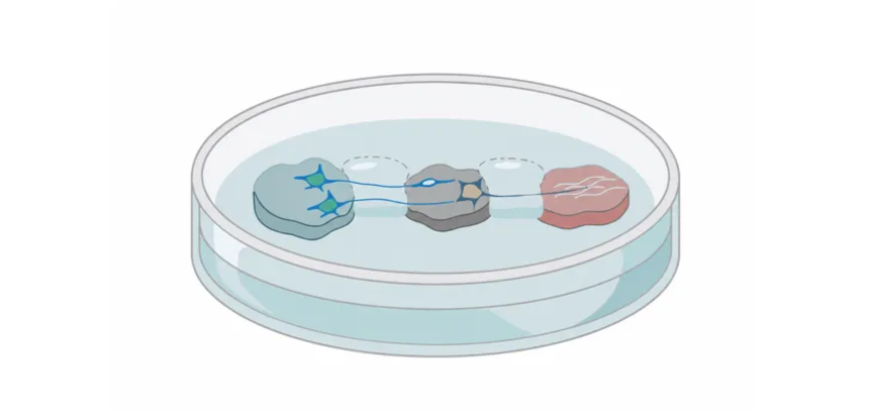
Now, in research published in Cell Reports, Dr Lakatos’s team has taken its research a step further, building a mini version of the connected human brain and spinal cord system in the lab by recreating these tissues using organoids.

In the human body, the brain and spinal cord tissues are distinct but connected by axons, so the researchers kept the brain and spinal cord organoids apart. They saw that nerve fibres from the brain tissue grew across the gap to connect to the spinal cord, forming a working circuit that could even cause tiny muscle clusters to contract.

By growing this human system in the dish for more than a year, they found that up until around day 150 – which corresponds to the mid-trimester of pregnancy – the axons were able to regrow after damage, but after this time, their growth was greatly impaired.

George Gibbons from the Department of Clinical Neurosciences at the University of Cambridge, the study’s first author, said: “Neurons taken from less mature organoids regrew long fibres after injury, but those from more mature organoids showed a sharp drop in their ability to regrow. In other words, poor regeneration is built into human neurons as they mature in the central nervous system.”

By analysing the gene expression – a sign of how active the genes are – in neurons that connect the brain and the spinal cord, they were able to identify a network of genes that acts as a ‘switch’ restricting the axon growth ability while the neurons mature to form connections (synapses). Amazingly, blocking key regulators of this network switched back on the ability of axons to grow.

The team then scanned a database of drug compounds to search for those that act on the genes in this network and identified as a candidate lynestrenol, a hormone drug licensed for managing certain menstrual disorders and as a contraceptive. When they tried this drug on damaged neurons, they found that it significantly boosted axon regrowth.

While scar tissue and inflammation may also restrict axon repair, exploring and tackling neuron-specific causes – the subject of this study – is very important. This is supported by evidence that axons of less mature neurons can grow through non-permissive environments that characterise injury sites.

Senior author Dr András Lakatos, who led the project at the Department of Clinical Neurosciences, said: “When the brain and spinal cord are damaged, the nerve fibres that carry movement signals from the brain to the spinal cord rarely grow back. That’s why paralysis is usually permanent. But we didn’t know exactly when the ability of axons to regenerate becomes limited. Our model provides a good indication that this block happens during development, and it can still be reversed after this point.

“Lynestrenol itself may not be the answer to spinal cord repair, but it shows us that, in principle, it should be possible to directly target human neurons and regenerate their axons. Although we still need to show that this strategy will also help to re-establish appropriate connections between the brain and spinal cord cells, this gives us hope that one day we may be able to treat conditions previously thought untreatable.”

Organoid models are an important way of understanding human biology. While animal models – for example, mice and rats – are useful for studying our biology as they share some similarities with humans, their differences ultimately limit what we can learn. Organoids grown from human stem cells can more closely mimic human biology.  

Dr Lakatos added: “Much of what we know about nerve regeneration comes from rodents, whose neurons behave differently from human neurons. Our sophisticated organoid models help bridge the knowledge gap from animal models to what we see in patients. They are also an important contribution to efforts to reduce the use of animals in research.”

Organoids, often referred to as ‘mini organs’, are being used increasingly to model human biology and disease. At the University of Cambridge alone, researchers use them to repair damaged liversunderstand Crohn’s disease in children, and model the early stages of pregnancy, among many other applications.

The research was funded by the UK Research and Innovation Medical Research Council and Spinal Research. Dr Lakatos has a long association with Spinal Research, from being a funded PhD student to now sitting on the charity's Grant Advisory Board Committee. 

Spinal Research Chief Executive Louisa McGinn said: “Today, we are entering a new era of hope and possibility for the 15million people worldwide living with a spinal cord injury.

“The next five years present an unprecedented opportunity to change what’s possible for people living with spinal cord injuries. Breakthrough therapies are nearing clinical reality and frontier technologies are creating bold new pathways toward repair and recovery.

"Spinal Research is committed to funding the most promising research and the best researchers around the world. The incredible work that Andras and his Group are doing at Cambridge shines a powerful light on what that can achieve and we’re delighted to support it.”

Researchers have found that when you recall and visualize something you have seen before—a key component of long-term memory—neurons in your visual system reactivate in a manner consistent with vision

Creative endeavors, like making art, writing music, or penning a poem, require the recall of memories to fuel imagination. Many other human behaviors, including problem solving, also rely on mental imagery to complete tasks, but little was known about how imagery works at the level of single neurons in the brain—until now.

Varun Wadia (PhD '23), a former graduate student in neurobiology at Caltech and now a postdoctoral scholar at Cedars-Sinai, and a team of scientists and physicians have found that many of the same neurons that are active when looking at an object are also active when imagining that object from memory. The findings could help develop defenses against diseases that cause memory loss, like Alzheimer's, and assist in building more efficient artificial intelligence platforms. A paper published April 9, 2026, in the journal Science outlines the researchers' process and findings.

"We were very interested in trying to understand the mechanisms of mental imagery because they permeate many interesting human behaviors," says Wadia, who is first author of the study and whose thesis work forms the core of the paper. "What we saw is that when you imagine something you've seen before, your visual system is being put into the state that it was in when you first looked at it."

The new study builds on work by Doris Tsao (BS '96), a professor in neurobiology at UC Berkeley who was a Caltech faculty member from 2009 to 2021 and is a senior author of the paper. Tsao, who served as Wadia's PhD advisor, studies the representations of visual objects in nonhuman primates. Her research has found the mechanism that the brain uses to represent facial identity and the mathematical system used by the brain to organize visual objects, among other discoveries.

The first step in the team's work was to compare how humans process objects in the brain with the framework that Tsao had found in nonhuman primates—called a distributed axis code—in which individual neurons encode a specific dimension, or axis, of object space. Wadia and Tsao collaborated with electrophysiologist and neuroscientist Ueli Rutishauser (PhD '08), who is a faculty associate in biology and biological engineering at Caltech and a faculty member at Cedars-Sinai where he directs the Center for Neural Science and Medicine. The researchers, along with Rutishauser's clinical colleagues at Cedars-Sinai, recorded neuron activity in patients with epilepsy who have electrodes temporarily implanted in their brains to monitor seizures.

The electrodes allowed the researchers to employ single-neuron recordings to document the activity of many individual neurons at the same time in a region called the ventral temporal cortex (VTC) that is critical to visual recognition and memory. This tool let the team examine what VTC neurons were doing when study participants looked at an object, such as a bird or a saxophone.

"It was very surprising how well the model for nonhuman primates mapped to and worked for humans," says Rutishauser, a senior author on the study. "It means this entire body of knowledge that has been developed over many years applies to the human brain, which is far from trivial."

Wadia and the team used neural activity to figure out how the neurons were representing viewed objects and then applied that knowledge to reconstruct the objects patients were viewing. They then had patients imagine a subset of the objects shown to them while recording activity from the same neurons. Roughly 40 percent of the neurons reactivated during this imagining phase and had similar responses as during vision, implying that both processes used the same distributed axis code. Furthermore, the responses were so strong that the researchers were then able to reconstruct the objects that people were imagining, a first for these types of studies.

"We did analysis to demonstrate that this reactivation is visual in nature and involves the same neurons, which suggests that we have a generative model in our heads," Wadia says. "That is an intriguing conclusion because it means we have a way to conceptualize how the nervous system implements creative tasks, whether it's making a song or a painting or imagining how to solve a problem in your head. This insight can serve as a hook into understanding all of those super interesting behaviors."

He says that having a mechanistic understanding of how creative and intelligent behaviors happen could help inform a more efficient way of developing artificial intelligence in computing systems. On the clinical side, Wadia and Rutishauser believe their findings represent the first step in decoding memory in the brain.

"If you really understand how memory works, then you can start to think about how we might consolidate memory or prevent it from being eroded by Alzheimer's and other diseases," Wadia says. "Tomorrow's clinical care is today's science project, and we are immensely grateful for the patients who were willing to participate in our work and understand that their participation could lead to benefits for others in the future."

The findings could also help reveal new solutions for mental illnesses such as schizophrenia. "There are many devastating conditions where people imagine things that don't exist and that has a negative effect on their well-being," Rutishauser explains. "Our work could have significant relevance in the field of psychiatry."

The team is also planning follow-up studies with additional data collected from their study participants to try and find where the trigger signal for reactivation is coming from and how different areas of the brain might be working together to implement imagination.

"This paper is really a good example of the kind of discoveries we can achieve when scientists and engineers at Caltech work closely together with clinicians and patients," Rutishauser says. "It's something that neither Caltech nor Cedars-Sinai could have done on its own."

The Science paper is titled "A shared code for perceiving and imagining objects in human ventral temporal cortex." Additional authors from Cedars-Sinai are neurologists Chrystal Reed, Jeffrey Chung, Lisa Bateman, and neurosurgeon Adam Mamelak. The authors also acknowledge Ralph Adolphs (PhD '93), Bren Professor of Psychology, Neuroscience, and Biology at Caltech, for his support of this study. Funding was provided by the National Institutes of Health's BRAIN Initiative, the National Institute of Mental Health, the Howard Hughes Medical Institute, the Simons Foundation Collaboration on the Global Brain, and the T&C Chen Center for Systems Neuroscience at Caltech.

Written by Katie Neith

https://www.caltech.edu/about/news/imagine-that-brain-uses-neurons-from-vision-system-when-forming-mental-imagery

Credit: Olivier Wyart ( www.headquarter.paris)


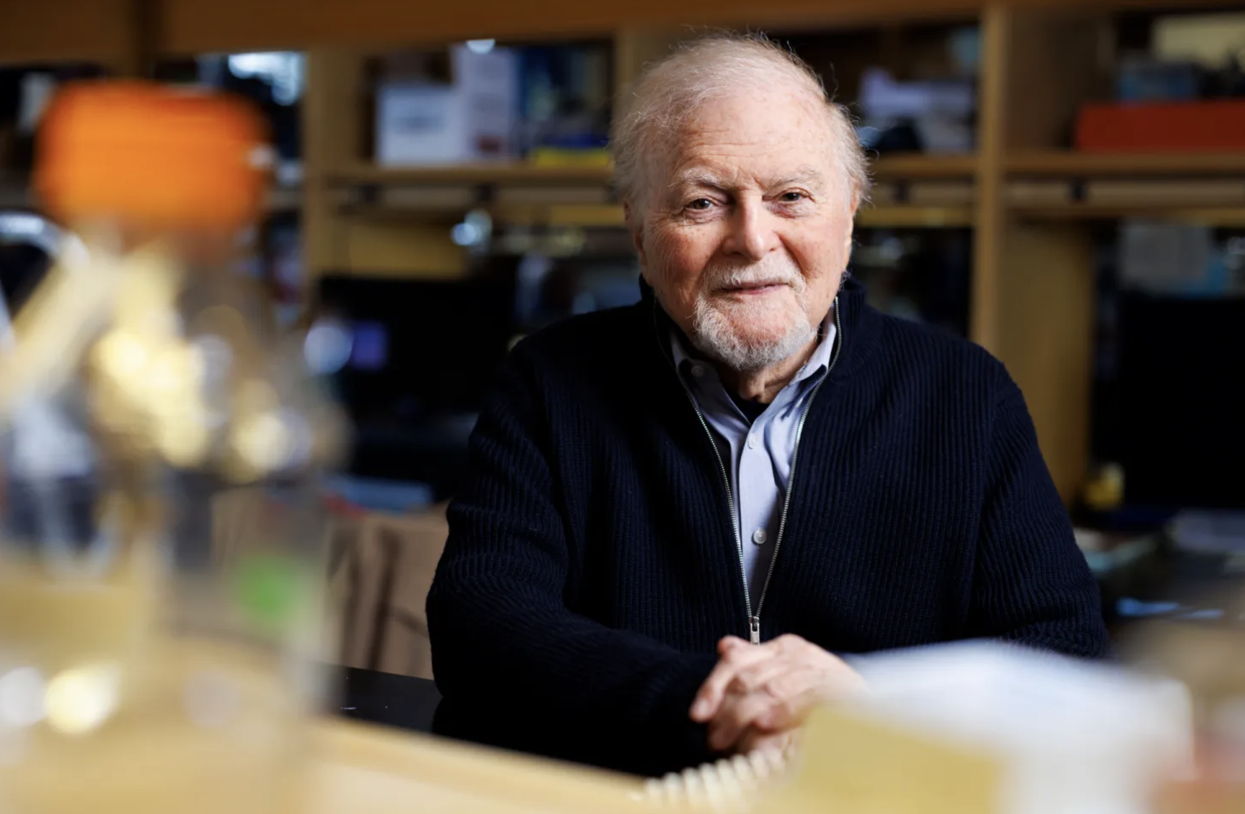
‘It’s not an imagined headache, and it’s not a mild condition,’ says Michael Moskowitz, Brain Prize recipient for his dogma-defying research

About 15 percent of people worldwide suffer from migraine, a neurological condition that can cause headache, nausea, visual disturbances, and sensitivity to light and sound. 

After stroke and neonatal brain injuries, migraine is the third-highest nerve-related cause of years lost to disability worldwide. 

The condition is also undertreated and poorly understood, says Michael A. Moskowitz, Harvard Medical School professor of neurology at Mass General. Moskowitz has made multiple discoveries that have revolutionized thinking about the condition, deepened knowledge about why some treatments work, and led to new treatments that are available and prescribed now. 

Moskowitz’s interest in neurology began early. At 14, he worked as a messenger in a hospital for patients with chronic neurological diseases near his family’s home in New York City, and was shocked by what he saw. “I could not comprehend how the brain could so easily betray the body,” he said. 

When he began his career in the early 1970s, migraine was still poorly understood. Images of the brains of patients came back totally normal: On paper, there was nothing wrong. 

“When I first started in the field, many people believed that migraine was a psychological problem,” said Moskowitz. “But it’s not an imagined headache, and it’s not a mild condition.” 

His first step, when he was a postdoctoral fellow and junior faculty in the Harvard-MIT Program in Health Sciences and Technology, an inter-institutional collaboration between Harvard University, Harvard Medical School, and MIT, was to dive into the literature. 

“I don’t know how many nickels I dropped in the Xerox machine at the Countway Library, but quite a few,” he said.

He found that no scientist had yet mapped the nerves carrying sensation from the circle of Willis, a network of arteries in the innermost layer of the meninges that supply blood to the brain. The brain itself doesn’t register pain, but the meninges, the brain’s three-layered covering, do. It seemed a promising place to start. 

So using a novel polymer-based technology developed in partnership with MIT chemical engineer Robert Langer, Moskowitz showed that nerve fibers that wrap around the circle of Willis travel back to the brain via the trigeminal nerve, which also carries sensation from the forehead, where headaches are often felt. His lab then found that these nerves contain and release neuropeptides, setting up a cascade that causes meningeal inflammation and other harmful effects. 

It was a stark departure from the previous belief about migraine, which was that the condition was purely caused by the dilation of blood vessels. 

In later research, Moskowitz demonstrated that classical migraine drugs called ergots and triptans acted in a completely different and unexpected way than had been assumed: Rather than constricting blood vessels, the drugs blocked those harmful neuropeptides from being released from nerve fibers in the first place. 

“That changed 100 years of dogma about how the ergots worked,” Moskowitz said. It also led to a new class of drugs that blocked neuropeptide release without vessel constriction In addition, it led to the development of drugs and antibodies that block the action of CGRP, a major neuropeptide in this pathway; those drugs are still in use today. Other neuropeptides discovered through his research are providing promising leads for future migraine therapies, said Moskowitz, who in 2021 received the Brain Prize for his contributions to migraine research. 

Building on the breakthrough, his lab began to look for the trigger that caused the release of peptides in the first place. They identified cortical spreading depression, a slow-moving tsunami of electrical and chemical changes in the brain. As the wave progresses, it can trigger migraine’s varied symptoms. For example, the migraine’s classic visual aura occurs as the chemical and electrical changes move through the brain’s visual cortex. 

Moskowitz’s research is also focused on the study of stroke and its potential neurovascular targets. In 2024, he and his chief collaborator, Matthias Nahrendorf, an HMS professor of radiology at Mass General, were awarded a Javits Award, a prestigious seven-year research grant, from the National Institutes of Health. Along with another collaborator, Charles Lin, an HMS professor of dermatology at Mass General, they are following up on new discoveries showing that the skull bone marrow and its blood-forming inflammatory cells contribute to the health of the meninges. The findings could impact a variety of neurological diseases, including stroke, Alzheimer’s disease, and multiple sclerosis. 

“My research has been 98 percent funded by the NIH over the course of my career,” Moskowitz said, expressing gratitude for the federal partnership that allowed bench-to-bedside medicine to flourish. “I can say with great confidence that if it weren’t for the NIH, we definitely wouldn’t have these new migraine drugs that block headaches.” 

Text: Sy Boles Harvard Staff writter

Veasey Conway/Harvard Staff Photographer

EU-backed researchers are pioneering a minimally invasive, graphene-based tool for detecting Alzheimer’s disease years before symptoms appear

The early detection of Alzheimer’s disease remains one of the most pressing challenges in modern neurology. With current approaches relying on expensive imaging techniques or invasive cerebrospinal fluid analysis, patients frequently end up being diagnosed at advanced stages, where treatment has limited effectiveness. The EU-funded 2D-BioPAD(opens in new window) project is addressing this critical gap by developing a point-of-care diagnostic tool that enables early detection through a simple, far less invasive blood test.

Graphene-based tech

The device relies on a sophisticated electrochemical biosensor constructed from graphene, a 2D material renowned for its exceptional strength and conductivity. By incorporating specific aptamers – synthetic molecules engineered to bind precisely to target proteins – the tool can simultaneously identify several biomarkers with a tiny drop of blood. This makes it possible to detect disease indicators at far lower concentrations than conventional methods.

Designed for use outside specialised hospitals, the system allows diagnosis to be made by general practitioners and at community health centres. The purpose is not to replace brain scans and spinal taps but to make earlier screening – and therefore earlier detection – possible, before symptoms appear and when treatments are more effective.

“There are good reasons for knowing you may go on to develop dementia,” comments Vincent Bouchiat, CEO and co-founder of French 2D-BioPAD project partner Grapheal, in an article(opens in new window) published on the website of ‘Horizon, The EU Research & Innovation Magazine’. “New Alzheimer’s medications show promise in delaying the progression of the disease, which is obviously a huge step forward.”

An AI helping hand

A defining feature of the 2D-BioPAD approach is its strategic integration of AI to overcome traditional limitations in biosensor development. The project is using machine learning not only for data analysis but also as a fundamental tool in the biosensor design phase. AI is being used to identify aptamer sequences for specific biomarker targets to help speed up aptamer selection, narrow down the pool of aptamer candidates and predict how well specific aptamers will function in the graphene-based device. AI models are also being used to explore improvements in the manufacturing of the biosensor. The initial focus will be on optimising its functionalisation using publicly available data and input from technology partners, with the potential to also improve sensitivity, defect levels and conductivity.

The advantages of the 2D-BioPAD tool are several. Firstly, and most importantly, the graphene sensor’s high sensitivity ensures that the disease can be detected before significant cognitive decline occurs, opening a critical window for treatment to take place when it could make a real difference. The minimally invasive nature of the blood test also reduces patient anxiety and discomfort. By being portable and easy to use, the device transforms Alzheimer’s screening from a complex, hospital-based procedure into a routine part of primary care, reducing the burden on healthcare systems and likely significantly lowering diagnostic costs.

“Early detection will only be realistic for people when it’s inexpensive for the health service, and painless and simple for the patient,” states Aristeidis Bakandritsos, a senior researcher at the Czech Advanced Technology and Research Institute, which is part of project coordinator Palacký University Olomouc in Czechia. With pilot studies underway in Germany, Greece and Finland, 2D-BioPAD (Supple Graphene Bio-Platform for point-of-care early detection and monitoring of Alzheimer’s Disease) promises to redefine the landscape of Alzheimer’s diagnosis, offering hope for earlier intervention and improved patient outcomes.

A fully implantable device that mimics the pancreas could dramatically ease life for people with type I diabetes

Type 1 diabetes is an autoimmune condition in which the pancreas can no longer secrete insulin. For millions of people, this translates into measuring glucose, monitoring carbohydrate consumption and injecting insulin on a daily basis.

Hybrid closed-loop systems have significantly improved glycaemic control and quality of life through continuous glucose monitoring and subcutaneous insulin pumps. However, subcutaneous delivery remains suboptimal because insulin absorption is relatively slow, requiring careful meal planning and frequent patient intervention.

A system for intraperitoneal delivery of insulin

The EU-funded FORGETDIABETES(opens in new window) project proposes a radically new therapeutic approach: a fully implantable bionic pancreas delivering insulin via the intraperitoneal route.

“Intraperitoneal delivery more closely resembles normal physiology, enabling insulin to act and clear more rapidly,” explains Claudio Cobelli, Emeritus Professor of Bioengineering at the University of Padova and project coordinator.

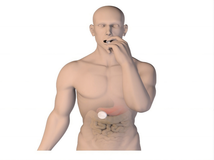
The system integrates three core components: a glucose sensor, a control algorithm and a pump connected to an internal insulin reservoir. A novel feature is the use of an oral insulin capsule(opens in new window) that patients must ingest once a week. These pills passively travel along the gastrointestinal tract until reaching the implanted device, where they refill the reservoir. This approach reduces the psycho-social burden associated with frequent injections

Importantly, the system uses novel biocompatible and immune-optimised coatings, guaranteeing long-term safety and stability. Continuous data transmission to the cloud ensures disease management.

Adaptive algorithms for precise glucose control

At the heart of the system lies an advanced control algorithm tailored to each patient. Maintaining blood glucose within the recommended target range requires precise insulin dosing, as too little can lead to hyperglycaemia and too much can cause hypoglycaemia. The adaptive algorithm continuously interprets sensor data and adjusts delivery accordingly.

Because intraperitoneal administration more closely mimics the natural pancreas(opens in new window), researchers hope it may eventually allow for narrower glucose targets and improved metabolic stability. The device is designed to respond rapidly to changes following meals or exercise, reducing the need for constant user input. Moreover, it is charged through wireless power transfer from a wearable patch outside the body.

Device testing and validation

The consortium has successfully tested the system in diabetes animal models. If successful, the technology could significantly reduce the numerous diabetes-related actions many patients currently perform each month, replacing them with only a few maintenance steps.

Ultimately, the ambition is to move closer to an automated, fully implantable artificial pancreas that relieves the daily burden of disease management. By combining physiological insulin delivery with intelligent control, FORGETDIABETES seeks to offer people with type I diabetes greater freedom, improved metabolic control and a substantially enhanced quality of life.

Beyond clinical performance, the project also addresses long-term reliability and scalability. According to Cobelli: “The next steps include additional animal studies, miniaturisation of device components as well as increasing the compatibility and durability of the implant according to European medical device standards.”

Long-term these will bring the FORGETDIABETES solution closer to clinical implementation, making life easier for people living with type I diabetes.

Image:  Padua University

Real-world data can reveal how patients truly recover after cancer. A European initiative combines clinical records and wearable data to improve care beyond the clinic

Recovering from breast cancer often involves managing a complex set of chronic conditions that persist long after primary treatment ends. However, a lot of clinical research still relies on snapshots from infrequent hospital visits, leaving critical gaps in understanding how patients function and feel in their everyday lives.

Clinical research has the potential to be transformed by tapping into real-world data(opens in new window), health and lifestyle data collected for each patient from electronic health records (EHR), digital registries, smartphones and wearable devices.

A 360° view of patient recovery

The EU-funded REBECCA(opens in new window) project brings together 12 partners from seven European countries and aims to exploit the novel potential offered by the continuous collection of real-world data.

To track patients’ functional, emotional and quality-of-life trajectories, the consortium developed a 360° Monitoring Platform, a suite of minimally obtrusive mobile tools offered to breast cancer survivors. The platform integrates EHRs and patient-reported outcome measures with passively collected digital biomarkers, including physical activity, heart-rate response, sleep patterns, eating habits and mobility trajectories.

“The goal was to improve patient care by studying the long-term effects of primary and adjuvant cancer treatments,” explains project coordinator Anastasios Delopoulos.

By aggregating and visualising these multimodal data streams, clinicians gain a far richer and more continuous understanding of patient recovery than is possible through routine follow-up visits alone. Instead of relying on appointments spaced months apart, healthcare professionals can observe recovery trends in near real time and receive automated alerts when abnormal patterns emerge.

Using AI to interpret real-world studies

The REBECCA platform has been deployed in six clinical studies across Norway, Spain, and Sweden, involving more than 650 participants. To interpret these rich datasets, REBECCA combined deep learning with AI-powered modelling. The models extract key information from real-world data and from the clinical trials, capturing key determinants of health and quality of life. Causal relationships among variables influencing breast cancer outcomes may also emerge.

“By linking treatments, behaviours, and functional outcomes, the models provide actionable insights that support informed clinical decision-making,” emphasises Delopoulos.

Early findings demonstrate that continuous monitoring can detect changes in activity, sleep or heart rate weeks before they would typically surface in routine care. These insights demonstrate the added value of real-world data in capturing aspects of recovery that matter most to patients but often remain invisible in standard clinical settings.

Building a foundation for future practice

Among REBECCA’s most significant achievements is the creation of a robust infrastructure for privacy-preserving, federated analysis of cross-country data. Just as important has been the project’s close collaboration with patient representatives, including the Swedish group AMAZONA, whose involvement helped ensure that the platform remains patient-centred and clinically relevant.

As the project concludes, REBECCA leaves behind a unique multimodal dataset and a scalable platform designed for continued use. Next steps include deploying the infrastructure in additional oncology clinics, extending it to other cancers and chronic diseases, and refining causal models by integrating behavioural and biological data. Through these efforts, REBECCA aims to support the wider adoption of real-world data as a cornerstone of clinical research and patient management across Europe.

Image: © www.thesmartcityjournal.com

Large new global study fuels growing concern over trend of increases in several types

Six cancer types are rising faster in younger adults than in those who are older in at least five countries, a new study of global cancer incidence shows, and two types — colorectal and uterine — are becoming both more common and more deadly among the young.

The massive study combed through data from two large cancer databases to better understand the recent rise of cases in adults under 50, a trend that belies the traditional understanding of the disease as one that disproportionately affects the elderly. Though still relatively rare among those in middle age and younger, the rising incidence of several cancer types in that cohort has raised concern among experts.

The work, published in November, painted a disturbing yet complex picture that varies globally according to cancer type, sex, and national context. The study examined cases that occurred between 2000 and 2017 and found 13 cancers on the rise in those under 50 in at least 10 countries, and six cancers — colorectal, cervical, pancreatic, prostate, kidney and multiple myeloma — rising faster in younger adults than in older adults in at least five.

The trends of both higher incidence and mortality in those under 50 occurred in fewer countries — in five for uterine cancer and, for colorectal cancer, three nations for females and five for males.

Colorectal cancer, particularly in North America, Europe and Oceania, drew particular attention from the authors, who said that 10 percent of global cases already occur in those under 50.

They cited estimates that, by 2030, colorectal cancer incidence in those ages 20 to 34 will rise by 90 percent, and, in those ages 35 to 49, by 46 percent.

The news is better, however, for late-onset colorectal cancer, with several countries showing incidence declining, likely in part due to screening programs that target older adults and detect precancerous growths early.

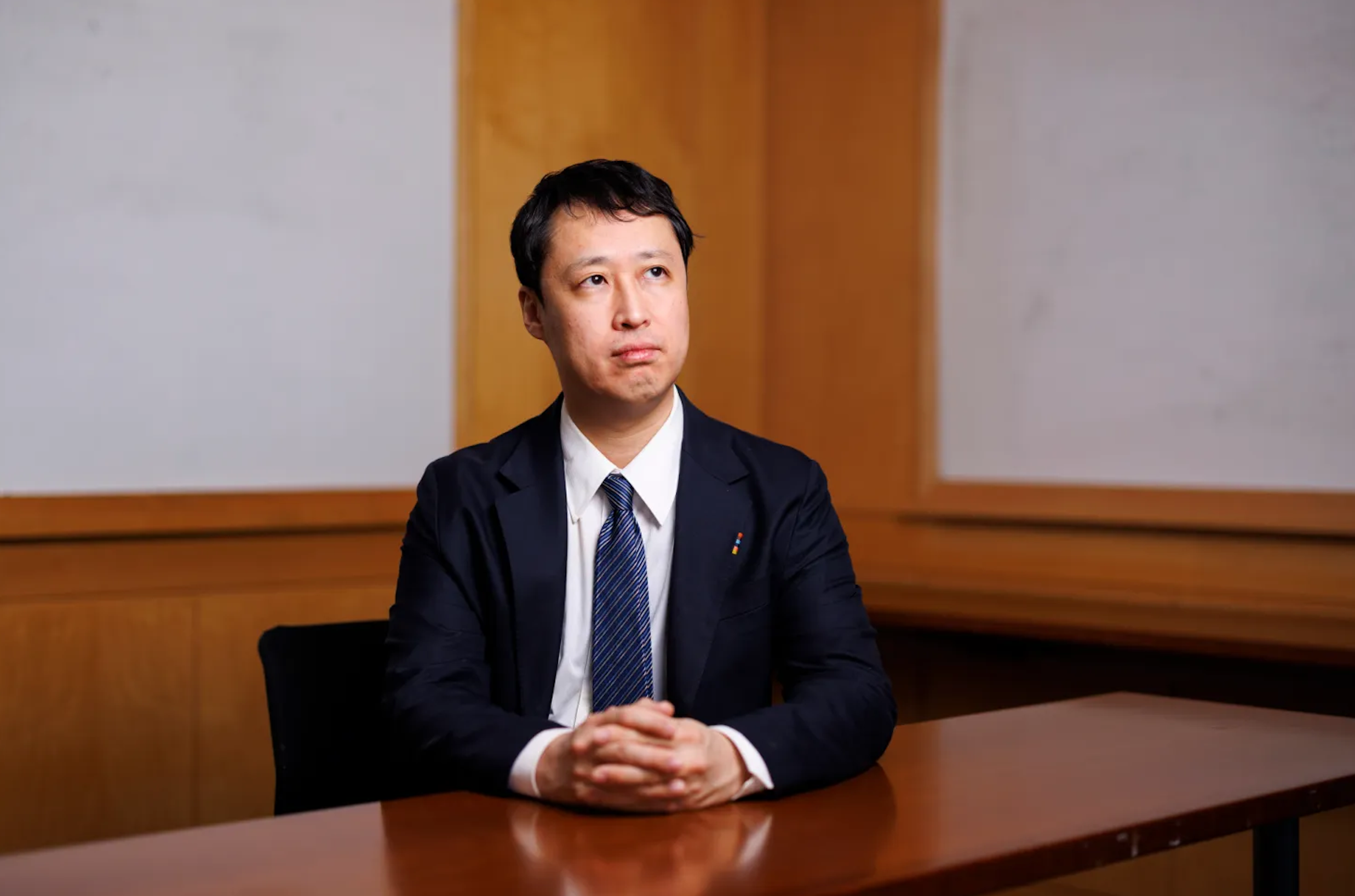
Tomotaka Ugai, an instructor in pathology at Harvard Medical School and Brigham and Women’s Hospital, a department associate in epidemiology at the Harvard T.H. Chan School of Public Health, and the study’s senior author, said though additional work is needed, the study does fill in some blanks in a concerning global cancer picture.

“There is a possibility that exposures like obesity or Western diet or sedentary lifestyle might be shifted toward younger populations.”

Despite the broad patterns seen, however, the study was limited by the fact that the databases used do not cover every nation. Missing are parts of Asia, Africa, and South and Central America.

Ugai said that a more detailed understanding of the young-onset cancer picture in every nation is needed to design interventions such as enhanced screening guidelines.

Some countries have already taken such steps

In the U.S., for example, screening guidelines have shifted younger in recent years. In 2021, the U.S. Preventive Services Task Force lowered the recommended age to begin screening for colorectal cancer from 50 to 45. Similarly, in 2024, it lowered the recommended age to begin breast cancer screening from 50 to 40.

In their results, in fact, the researchers, from the U.S., Japan, and South Korea, detected evidence of already improved screening procedures for some cancers, which the authors said may be one factor in rising incidence rates.

In those cancers, including thyroid, prostate, and non-melanoma skin cancers, there was no increase in mortality despite rising incidence, likely an indication that improved screening is detecting more cases early enough that they are treatable, or that are clinically insignificant and may have been missed before.

The analysis, published in the journal Military Medical Research and supported by private funding, also provided clues about the reason for the rise in young-onset cancers. The increase in cancer among younger adults is strongly tied to rising rates of obesity, the authors said, and is climbing most rapidly in wealthy nations.

Future work, Ugai said, will involve international collaborations to better understand what’s going on in these cases. They’ll also augment data from large-scale studies with analyses of individual tumor tissues in an effort to better understand mechanisms in tumor biology and microenvironments that might be at work.

“Many studies, including our studies, indicate that the incidence of early onset cancer has been increasing in many parts of the world,” Ugai said. “But that doesn’t mean that many cancer types have been increasing similarly everywhere. All over the world, the situation is very, very different, so we need to know more about what’s happening now, what are the risk factors, and how to prevent these cancers.”

Text: Alvin Powell /Harvard Staff Write

Photo: Tomotaka Ugai, senior author of a new global cancer study.(Veasey Conway/Harvard Staff Photographer)

Page 1 of 3
We use cookies

We use cookies on our website. Some of them are essential for the operation of the site, while others help us to improve this site and the user experience (tracking cookies). You can decide for yourself whether you want to allow cookies or not. Please note that if you reject them, you may not be able to use all the functionalities of the site.